 Hi there, it’s been a while since I last wrote. I’ve been quite busy and I haven’t really had the time or energy to update the blog. I have a little bad conscience because I just disappeared from the blog without saying anything. But now I’m back with an update for those of you who are interested in what has happened in my life the past year. Because it’s been an eventful year and I’m happier than ever.
Do you remember that I was joking about that there must be someone out there for me? Someone who likes casino, Sinatra, sports, junk food and traveling? I finally found her. She does not meet all the criteria, for example she does not like Frank Sinatra or junk food, but she is perfect anyway. We actually met at a casino and it was love at first sight, at least for me. After a few dates we decided to go to Vegas together and it was an experience out of the ordinary.
If you ever get the chance to go there – take it! We had great fun and went into the tourist role to 100%. We looked at the famous Las Vegas sign, took a limousine ride along the strip and checked out Downtown where Las Vegas once began to emerge. Of course there were also some casino visits and we tested both roulette, black jack and different slots. Although we tried to do as much as possible during the trip, we also enjoyed some relaxation at the hotel pool. I really miss the pool and wish I could have brought it home with me.
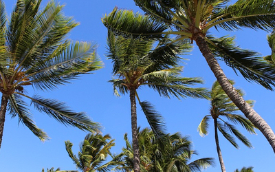
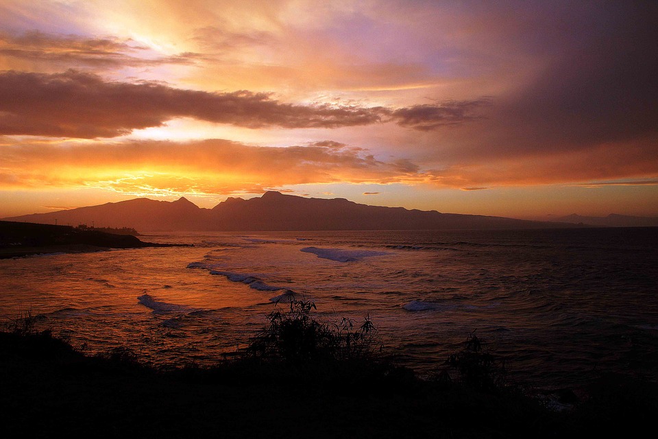
After the Vegas trip, I was sure she was the right one so I proposed. Fortunately, she said yes. So now I’m happily engaged and plans the next big trip that will go to Hawaii. As you know, both me and the children want to go back to Hawaii and now it finally looks like it will happen. Longs very much and I think it will great.
Until we travel, however, I will work as usual. The holidays are over and it’s evident in the bookings.
 I must say that it´s hard to run a blog. I tend to loose my motivation real fast. I have been thinking about this and decided to give it another try. So, what´s up with me?
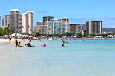 Hi again guys! Sorry for being so late with my next update, but I’ve been so busy with work and working out (yes, I now work out) so the site had to suffer a bit. However, now I’m back to the every day life and I feel like I have the time to tell you a bit about my trip to Hawaii. I went with my daughter and we had a great time! Again, thank you all for the wonderful tips!